
Reducing Bacterial Bioburden in Infected Wounds with Vacuum Assisted Closure and a New Silver Dressing -- A Pilot Study
Abstract and Introduction
Abstract
The use of silver is increasing rapidly in the management of infected wounds and wounds at risk for infection. This 5-patient case series reviews the results of a pilot study designed to determine efficacy and safety of negative pressure wound therapy (NPWT; V.A.C.® Therapy™, KCI, San Antonio, Tex) with the new silver foam dressing (V.A.C.® GranuFoam® Silver™ Dressing, KCI). Data on wound progression and the primary endpoints-time to clear infection, time to wound closure, and time to discharge from hospital-are detailed in this manuscript. For the 5 patients treated with NPWT and the silver foam dressing, the mean time for treatment was 13.00 ± 3.39 days and the mean time to patient discharge was 16.00 ± 6.63 days. Wounds were clear of clinical signs of infection in 7.00 ± 1.58 days and closed in 19.20 ± 8.76 days.
Introduction
Infections complicate the treatment of wounds and impede the healing process by damaging tissue, reducing wound tensile strength, and inducing an undesirable inflammatory response.[1,2] Increased bacterial burden in a wound increases the metabolic requirements of the tissues, stimulates a proinflammatory environment, and encourages the in-migration of monocytes, macrophages, and leukocytes-all of which can negatively impact wound healing.
Bacteria can also secrete harmful cytokines, which can lead to direct vasoconstriction and decreased blood flow to the wound.[3] Thus, controlling or preventing infections is essential in order for the normal wound healing process to occur. In addition, reduction of infection can also yield significant overall cost savings in healthcare, as infections lead to thousands of dollars in excess medical charges and lengthened hospital stays every year.[4]
Negative pressure wound therapy (NPWT; V.A.C.® Therapy™, KCI, San Antonio, Tex) was introduced by Argenta and Morykwas in 1995 to aid in the healing process by removing interstitial fluid and infectious materials through the application of localized negative pressure to the wound.[5,6] The negative pressure applied to the foam of the NPWT system induces undulations in the wound surface, thereby stretching cells and promoting cell division and the proliferation of reparative granulation tissue.[7] Scientific studies have also demonstrated that the therapy promotes blood flow.[6,8] In clinical practice, the authors have found that NPWT can enhance the reliability of surgical procedures and have incorporated its use in a wide variety of applications, such as acute traumatic wounds, chronic wounds, dehisced wounds, degloving injuries, burns, pre- and post-skin graft applications and flap procedures, and in conjunction with skin substitutes.[9-17]
Ionic silver has long been recognized as an effective antimicrobial agent used against a broad spectrum of pathogens and is considered to be biocompatible with mammalian tissue.[18-23] The authors hypothesized that combining the antimicrobial activity of silver with the proven clinical benefits of NPWT would effectively reduce bacterial bioburden in wounds. For this pilot study, NPWT was applied with the new microbonded silver foam dressings (V.A.C.® GranuFoam® Silver™ Dressing, KCI) designed specifically to be used with this NPWT system.
Patients and Methods
A prospective case series of 5 consecutive patients with infected wounds treated with NPWT and the silver foam dressing was conducted. Each patient met the following inclusion criteria: at least age 18 with complex, open, infected wounds. Patient age ranged from 26 to 49 years. Three men and 2 women were treated. Four wounds resulted from open fractures sustained during motor vehicle accidents, and 1 wound resulted from a surgical abdominal wound dehiscence. The total area of the wounds ranged from 5 cm2 to 100 cm2, and the mean wound area was 43.00 cm2 ± 42.22 cm2 ( Table 1 and Table 2 ).
All wounds were sharply debrided of nonviable tissue before NPWT was applied. For all patients, a swab of soft tissue was taken and submitted on Day 1 of hospital admission for qualitative culture analysis. For Patient 1, the silver foam dressing was applied on hospital Day 30 following 30 days of NPWT with the standard foam dressing. For the remaining 4 patients, NPWT with the silver foam dressing was initiated as a first line therapy upon admission, following initial surgical debridement.
The US Food and Drug Administration-approved silver foam dressing used in this case series is an open-celled, reticulated polyurethane foam that is microbonded with metallic silver via a proprietary metalization process (Figure 1). The silver foam dressing was placed in the entire wound cavity and covered with an adhesive drape to create an airtight seal. A 1-2 cm round hole was cut into the drape. A T.R.A.C.® pad with tubing was applied directly over the hole and connected to a fluid collection canister, which is contained within the computer-controlled NPWT device.
 | Figure 1. Ionic silver is microbonded to the dressing in such a way that the foam retains the same porosity and structure of traditional foam dressing to simulate the same micromechanical effects. |
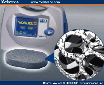
Figure 1.
No interface or adjunctive antimicrobial dressings were used with the silver foam dressing. This allowed for direct and complete contact of the dressing with the wound bed. For each wound, the NPWT device was programmed to deliver -125 mmHg continuous pressure, with dressing changes every other day. When the wounds were granulated to the surface and could be closed primarily or via secondary intention NPWT with the silver foam dressing was discontinued. Time to wound closure was defined by complete closure of the wound either by primary or secondary intention. Wound progression data and the primary endpoints—time to clear infection, time to wound closure, and time to discharge from hospital—were documented and recorded for all 5 patients ( Table 1 ). Age, wound area, history of diabetes, and albumin levels were also recorded ( Table 2 ).
Descriptive statistics and statistical analyses were calculated using SPSS 12.0 (SPSS Inc, Chicago, Ill) to determine demographic and result endpoints for the silver foam dressing-treated patients. All categorical variables are expressed as frequency and percentages, while continuous variables are expressed as the mean ± standard deviation, unless otherwise stated.
Results
No complications were experienced during the application of the silver foam dressings for any of the 5 patients. All wounds progressed to the point of primary or secondary closure. The times to infection clearance and wound closure were 7.00 ± 1.58 and 19.20 ± 8.76 days, respectively. The mean number of days required for treatment was 13.00 ± 3.39 days, and healthy granulation tissue was present in 5.50 ± 2.08 days. Mean time to patient discharge was 16.00 ± 6.63 days ( Table 1 and Table 3 ).
Case Reports
Case 1. A 49-year-old man was admitted with an infected, open, left ankle wound. Six months prior to this admission, the patient sustained a distal tibial fracture, which was plated during surgery immediately following the incident. Upon this admission, the wound cultured positive for Staphylococcus and vancomycin-resistant Enterococci (VRE). Antibiotic therapy was initiated immediately for a total of 1 week. The infected plate was removed, and the wound was surgically debrided. The resulting wound area was 100 cm2 with exposed bone and tendon.
On Day 3 post surgical debridement and removal of the plate (Figure 2A), NPWT was initiated with the traditional foam dressing at -125 mmHg continuous pressure. Screw holes in exposed bone are visible in Figure 2A. On NPWT Day 22, the wound was 90% granulated (Figure 2B). However, on Day 30, the wound still cultured positive for VRE, which prompted a switch to treatment with the silver foam dressing (Figure 2C).
 | Figure 2. Case 1: A) wound post-debridement; B) Day 22 of NPWT with standard foam dressing; C) Day 30 silver foam dressing placed in wound cavity; D) Day 35 of NPWT, after 5 days of NPWT with the silver foam dressing; E) split-thickness skin graft placed on Day 39, after 9 days of NPWT with the silver foam dressing; F) 3-month follow-up post discontinuation of NPWT. |
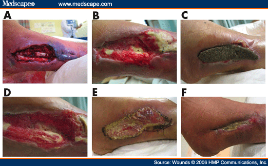
Figure 2.
On Day 35 of NPWT including 5 days with the silver foam dressing, the wound cultured negative for all clinical infection, including VRE (Figure 2D). Two days later, the wound was grafted with a bilayer matrix dermal regeneration template (Integra™, Integra LifeSciences Corporation, Plainsboro, NJ) on Day 37. The patient then underwent a split-thickness skin graft (STSG) on Day 39 and was discharged home (Figure 2E). The silver foam dressing was used on top of both the bilayer matrix template and the STSG.[14,16] A nonadherent dressing was placed between the silver foam and the skin grafts per recommended protocol.[24,25] On Day 42, NPWT was discontinued, and the STSG had 100% take without any sloughing or signs of infection. The wound remained closed at the 3-month follow-up visit (Figure 2F).
Case 2. A 47-year-old man sustained an ankle fracture 2 years prior to admission and presented with a necrotic wound over the prior surgical site. The wound culture returned positive for Staphylococcus, Enterococcus, Pseudomonas, Enterobacter, and Klebsiella. Further assessment revealed chronic osteomyelitis, which had been present and treated for 2 years. Upon admission, 6 weeks of antibiotic therapy were prescribed to treat the chronic osteomyelitis. The wound was surgically debrided, and the resulting wound measured 75 cm2 (Figure 3A). Immediately following surgical debridement, NPWT was initiated with the silver foam dressing at -125 mmHg continuous pressure.
 | Figure 3. Case 2: A) wound at admission, post-debridement; B) wound on silver foam dressing Day 12; C) wound on Day 15 of silver foam dressing (dressing switched to traditional foam dressing); D) wound on NPWT Day 22; E) 27 wound fully granulated to surface, NPWT discontinued, and patient discharge home; F) wound remains closed at 2-month follow-up post discontinuation of silver foam dressing. |
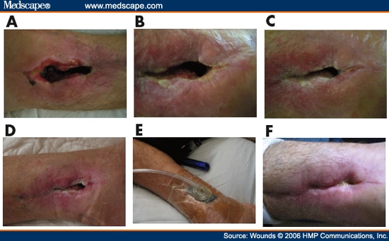
Figure 3.
On Day 6 of using the silver foam dressing, healthy granulation tissue was observed, and the wound cultured negative for all bacterial organisms. On Day 12, the wound was considerably smaller and covered with granulation buds (Figure 3B). On Day 15, the dressing was switched to traditional foam dressing (Figure 3C). The wound was further contracted on Day 22 (Figure 3D). Negative pressure wound therapy with the standard foam dressing was applied until Day 27 (Figure 3E), at which time the wound was fully granulated to the surface, and the therapy was discontinued. The patient was discharged home with a nonadherent dressing over the wound to optimize re-epithelization. The closed wound at 2-month follow-up post discontinuation of NPWT with the silver foam dressing is shown in Figure 3F.
Case 3. A 42-year-old man sustained a pilon fracture 2 years prior to admission and presented with a chronic open ankle wound. Due to infection, hardware had been removed 3 months prior to presentation. A culture returned positive for VRE. The total area of the wound after debridement was 10 cm2 (Figure 4A). Immediately following debridement, NPWT was initiated with the silver foam dressing at -125 mmHg continuous pressure (Figure 4B). Healthy granulation tissue was observed on Day 3 (Figure 4C), and the wound was further contracted on Day 5 (Figure 4D). On Day 9, the wound culture returned negative for VRE infection (Figure 4E). No systemic antibiotics were used during the length of therapy for this patient. Negative pressure wound therapy was applied for 15 days with the silver foam dressing at which point the wound was closed and the patient was discharged home (Figure 4F).
 | Figure 4. Case 3: A) wound at admission, post-debridement; B) Day 1 of silver foam dressing; C) Day 3 of silver foam dressing; E) Day 9 of silver foam dressing, wound cleared of infection; F) Day 15, patient is discharged with closed wound. |
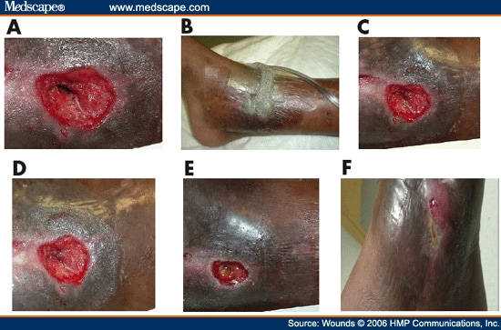
Figure 4.
Case 4. A 26-year-old woman sustained a proximal ulnar fracture from a motor vehicle accident. The patient was admitted with an infected, open ulnar fracture with exposed bone 5 days following the accident. The total area of the wound after debridement was 25 cm2 (Figure 5A). A wound culture returned positive for Staphylococcus aureus and Enterococcus. Negative pressure wound therapy was initiated with the silver foam dressing at –125 mmHg continuous pressure (Figure 5B). The silver foam dressing was applied for 14 days. No systemic antibiotics were used during the length of NPWT for this patient. The wound cultured negative for infection on Day 8. On Day 14, the wound was fully granulated and the patient was discharged for a STSG at another facility (Figure 5C).
 | Figure 5. Case 4: A) open ulnar fracture with exposed bone at hospital admission; B) silver foam dressing in place; C) Day 14 of NPWT with silver foam dressing, patient is discharged to another facility for skin graft. |
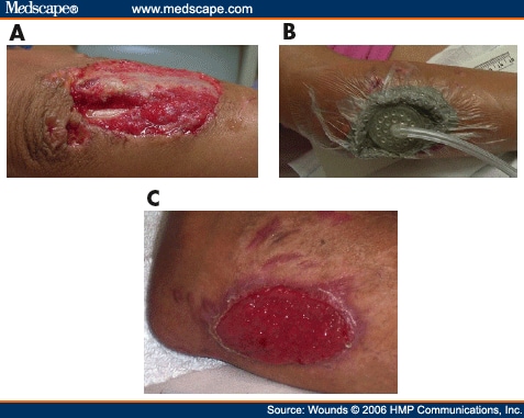
Figure 5.
Case 5. A 45-year-old woman with a history of type II diabetes mellitus, coronary artery disease, and obesity (body mass index of 56) was taken to the operating room for an open appendectomy and discharged home on postoperative Day 2. The patient subsequently presented to the emergency room on postoperative Day 5 with purulent drainage from the wound. She was admitted, and the wound was debrided. The total area of the wound after debridement was 5 cm2. A wound culture returned positive for Staphylococcus aureus and Enterococcus, at which time NPWT was initiated with the silver foam dressing at -125 mmHg continuous pressure. Negative pressure wound therapy was applied for 14 days with the silver foam dressing. No systemic antibiotics were used during the length of therapy for this patient. The wound cultured negative for infection on Day 7. On Day 14, the wound was fully granulated to the surface and contracted. Negative pressure wound therapy was discontinued and the patient was discharged home with antibiotic ointment and gauze. Complete re-epithelized closure was achieved on Day 20.
Discussion
From the present experience, the authors found the NPWT with the silver foam dressing to be a safe and efficacious adjunct in reducing bacterial bioburden in these wounds. The initial treatment results were noteworthy and showed a marked reduction in infection, wound closure time, and hospital stay over the institution's historically employed use of moist wound therapy for the management of infected wounds. The sample size of this prospective case series of 5 consecutive patients treated with the silver foam dressing is relatively small and was meant to minimize patient risk.
The authors have found the new silver foam dressing to be most useful in cases of complex, colonized, or infected wounds post debridement, as well as for acute traumatic wounds for reduction of bacterial bioburden. It is a preferred dressing to help reduce the risk of recurrent infection in patients who have severe comorbidities and a history of chronic wound colonization. The authors have also observed good results when the silver foam dressing is placed over a STSG, as the dressing has the necessary antimicrobial coverage in addition to the bolstering effect of NPWT. Conversely, the authors would indicate the traditional foam dressing instead of the silver foam dressing for wounds that are clear of infection or have low risk of recurrent infection.
In the past, the authors have used other silver antimicrobial dressings in combination with the traditional foam dressing with mixed results. Some silver dressings led to adverse reactions in wound healing, and other dressings have not provided the antimicrobial coverage needed to decrease the bacterial bioburden. In addition, some of the silver dressings do not have the wound conformability needed to eliminate areas of noncontact, which may allow bacteria to proliferate in those areas.[26] It has also been shown that negative pressure can be affected when an interface layer is used between the foam dressing and the wound.[27]
Use of the silver foam dressing in conjunction with NPWT offers a sufficient antimicrobial effect and a good bridge to wound healing. This may be due to a combination of factors including nontoxic levels of silver release (2-3 parts per million [ppm]), optimal wound conformability, nontraumatic exudate removal, and the proven clinical benefits of NPWT. In addition, the silver foam dressing is "user-friendly," as it does not require adjunctive antimicrobial dressings beneath the foam.
In each of the 5 cases, use of the silver dressing was continued past the point of infection clearance to act as a barrier to further bacterial penetration.[2] There were no incidences of recurrent infection. In 2 of the 5 cases, systemic infection was present, and intravenous antibiotics were administered to treat the invasive infection. In these situations, literature supports that topical antibiotic agents alone are not sufficient to reduce infection, and culture-specific systemic antibiotics should be prescribed.[3] An additional risk for these 2 cases was insufficient systemic antibiotic delivery due to an ischemic lower extremity. In these instances, the silver foam dressing provided effective topical antimicrobial action in addition to the systemic antibiotics. Systemic antibiotics were not prescribed in the remaining 3 cases, as the presence of topical infection only was suspected. Bacterial bioburden was reduced and wound closure was achieved without the need for systemic antibiotics.
The resurgence of interest in silver products for wound care stems from the increase in the level of bacterial resistance to traditional antibiotics. For example, rates of methicillin-resistant Staphylococcus aureus (MRSA) increased steadily over the past decade from approximately 30% in 1989 to approximately 40% in 1997 among intensive care unit patients.[28] Unlike traditional antibiotics, ionic silver has multiple mechanisms of action, such as inhibiting cellular respiration, denaturing nucleic acids, and altering cellular membrane permeability.[29,30] An adequate concentration of silver coupled with its various mechanisms of action make it difficult for microorganisms to develop resistance to silver because they would have to undergo several mutations in order to develop defense mechanisms against silver's multi-pronged attack.[3]
However, potential resistance to silver should be a consideration, and the adequate level of silver required for bioburden efficacy remains a controversial and complex issue. Some investigators believe low levels are enough, and others support the view that a higher level of silver is necessary for sufficient bacterial kill.[29-31] There is no global standard for elution studies, and the literature varies drastically, from 1.0 ppm to more than 36 ppm, in the suggested requirements for the proper kill levels.[31] A key confounding factor is that all pathogens have multiple strains, and different strains require different levels of silver for kill. There are also significant differences in methodology of measure, which complicate data interpretation of silver levels between various silver dressings.
Another important factor in measuring silver delivery is the ability of the dressing to conform to the wound site. Recent research indicates that the ability of a silver-containing dressing to conform to the contours of a wound is important to reduce areas of noncontact where bacteria may proliferate.[26] The silver effect was evenly distributed to the entire wound surface with the silver foam dressing.
Scientific research has determined that the application of NPWT with a traditional foam dressing induces micromechanical effects at the dressing-to-tissue interface, resulting in tissue undulations and cellular microdeformation.[32] The silver is microbonded to the silver foam dressing in such a way that the foam retains the same porosity and structure of the traditional foam dressing to simulate the same micromechanical effects (Figure 1). These localized micromechanical forces are thought to be a primary driver in cellular proliferation, angiogenesis, and stimulation of growth factors.[7,32] Therefore, interface layers were not used between the wound and the silver foam dressing, except in cases of protecting exposed tendon or a split-thickness skin graft, in order to maximize the effectiveness of the NPWT.
Some authors have suggested that higher levels of silver delivered by agents, such as nanocrystalline silver, can be harmful to viable cells. This toxicity of silver is supported in several in-vitro studies.[33,34] However, this is contrary to what the authors experienced in the clinic with this dressing. In each of the 5 cases in this series, use of the silver foam dressing was continued beyond the point of infection clearance because of consistently observed progression toward closure with the silver dressing. In 1 case, the silver foam dressing was applied over a split-thickness skin graft with the goal of bioburden reduction and enhanced graft take. The result was 100% graft take without sloughing or signs of infection. Demling and DeSanti[35] published similar findings in a controlled study that showed a moistened silver delivery system placed over a meshed skin graft on an excised burn wound that significantly increased the rate of re-epithelization compared to a standard antibiotic bolster dressing.
A recent study sheds an interesting light on silver-containing dressings and their variable effects on cells. Cochrane et al[36] used a fibroblast-seeded collagen gel model to evaluate the effect of 7 different silver-containing antimicrobial dressings on fibroblast contraction and viability. Of 7 dressings, 3 demonstrated less than 20% cell viability after 96 hours, compared to approximately 70% viability for the remaining 4 dressings. The authors concluded that silver dressing selection should be based on the overall characteristics of a dressing, including its antimicrobial, fluid-handling, physical, and chemical properties.
Numerous peer-reviewed publications support positive wound management aspects of nanocrystalline silver.[35-38] Wright et al[38] noted reduced levels of matrix metalloproteinases and a higher frequency of apoptosis in a porcine model of contaminated wounds treated with silver. The results suggest that nanocrystalline silver may play a role in altering or compressing the inflammatory events in wounds and facilitating the early phases of wound healing.
Conclusion
In this small, pilot study, NPWT with the new silver foam dressing demonstrated a reduction in the mean time to infection clearance, wound closure, and hospital discharge compared to the authors' previous moist wound care therapy. By incorporating the new silver foam dressing into practice, the authors have been able to reduce bacterial bioburden in chronic and acute wounds, while allowing the wounds to progress to healing with a healthy granulating wound bed. This improved rate of wound healing has allowed the use of simpler procedures for closure and aversion of more complex procedures, such as free flaps, and their related expenses.[39] Anecdotally, the authors have also noted a reduction in hospital length of stay for the patients treated with this combination therapy, which has yielded hospital savings in addition to the overall enhancement of patient quality of life. Further studies with larger patient samples hold promise for comparable results of this novel combination wound therapy.
Tidak ada komentar:
Posting Komentar